 |
| mammography |
Mammography is specialized medical imaging that uses a low-dose x-ray system to see inside the breasts. A mammography exam, called a mammogram, aids in the early detection and diagnosis of breast diseases in women.
An x-ray (radiograph) is a noninvasive medical test that helps physicians diagnose and treat medical conditions. Imaging with x-rays involves exposing a part of the body to a small dose of ionizing radiation to produce pictures of the inside of the body. X-rays are the oldest and most frequently used form of medical imaging.
Three recent advances in mammography include digital mammography, computer-aided detection and breast tomosynthesis.
Digital mammography
It also called full-field digital mammography (FFDM), is a mammography system in which the x-ray film is replaced by electronics that convert x-rays into mammographic pictures of the breast. These systems are similar to those found in digital cameras and their efficiency enables better pictures with a lower radiation dose. These images of the breast are transferred to a computer for review by the radiologist and for long term storage. The patient’s experience during a digital mammogram is similar to having a conventional film mammogram.
It also called full-field digital mammography (FFDM), is a mammography system in which the x-ray film is replaced by electronics that convert x-rays into mammographic pictures of the breast. These systems are similar to those found in digital cameras and their efficiency enables better pictures with a lower radiation dose. These images of the breast are transferred to a computer for review by the radiologist and for long term storage. The patient’s experience during a digital mammogram is similar to having a conventional film mammogram.
Computer-aided detection: (CAD) systems search digitized mammographic images for abnormal areas of density, mass, or calcification that may indicate the presence of cancer. The CAD system highlights these areas on the images, alerting the radiologist to carefully assess this area.
Breast tomosynthesis: It is also called three dimensional (3-D) mammography and digital breast tomosynthesis(DBT), is an advanced form of breast imaging where multiple images of the breast from different angles are captured and reconstructed ("synthesized") into a three-dimensional image set. In this way, 3-D breast imaging is similar to computed tomography (CT) imaging in which a series of thin "slices" are assembled together to create a 3-D reconstruction of the body.
What does the equipment look like?
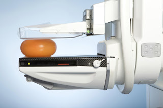
A mammography unit is a rectangular box that houses the tube in which x-rays are produced. The unit is used exclusively for x-ray exams of the breast, with special accessories that allow only the breast to be exposed to the x-rays. Attached to the unit is a device that holds and compresses the breast and positions it so images can be obtained at different angles.
A mammography unit is a rectangular box that houses the tube in which x-rays are produced. The unit is used exclusively for x-ray exams of the breast, with special accessories that allow only the breast to be exposed to the x-rays. Attached to the unit is a device that holds and compresses the breast and positions it so images can be obtained at different angles.
Breast tomosynthesis is performed using digital mammography units, but not all digital mammography machines are equipped to perform tomosynthesis imaging.
Benefits
🍄Imaging of the breast improves a physician's ability to detect small tumors. When cancers are small, the woman has more treatment options.
🍄The use of screening mammography increases the detection of small abnormal tissue growths confined to the milk ducts in the breast, called ductal carcinoma in situ (DCIS).
These early tumors cannot harm patients if they are removed at this stage and mammography is an excellent way to detect these tumors. It is also useful for detecting all types of breast cancer, including invasive ductal and invasive lobular cancer.
🍄No radiation remains in a patient's body after an x-ray examination.
🍄X-rays usually have no side effects in the typical diagnostic range for this exam.
Risks
📌There is always a slight chance of cancer from excessive exposure to radiation. However, the benefit of an accurate diagnosis far outweighs the risk.
📌The effective radiation dose for this procedure varies. See the Safety page for more information about radiation dose.
📌False Positive Mammograms, Five percent to 15 percent of screening mammograms require more testing such as additional mammograms or ultrasound. Most of these tests turn out to be normal. If there is an abnormal finding, a follow-up or biopsy may have to be performed. Most of the biopsies confirm that no cancer was present. It is estimated that a woman who has yearly mammograms between ages 40 and 49 has about a 30 percent chance of having a false-positive mammogram at some point in that decade and about a 7 percent to 8 percent chance of having a breast biopsy within the 10-year period.
📌Women should always inform their physician or x-ray technologist if there is any possibility that they are pregnant. See the Safety page for more information about pregnancy and x-rays
very nicely have explained the meaning for Mammography, thanks for sharing the information,if looking for more information related to Biomedical Engineering, visit: Biomedical Engineering Subjects
ReplyDeletegreat blog… thanks for sharing detailed information about mammography machine with its benefits.
ReplyDeleteThanks for sharing the benefits of Mammography Machine
ReplyDeleteThe explanation of mammogram machines will create the awareness about taking mammogram scans. We Radolabs Scan Centre Chennai primary motto is to provide affordable diagnostics.
ReplyDeleteExcellent article; many thanks for informing us. It's been extremely helpful. Keep sharing, please. If you want to learn more about the reasonable finest health package, please pick the link.
ReplyDeleteMaster Health Checkup in Coimbatore