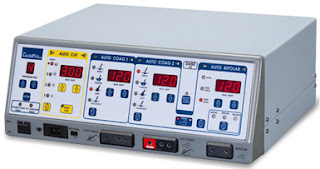
 |
| surgical diathermy |
The
purpose of this guideline is to provide guidance about surgical
diathermy.
Electrosurgical
units (diathermy machines) were first introduced during the early
twentieth century to facilitate haemostasis and/or the cutting of
tissue during surgical procedures
This
is achieved by passing normal electrical current via the diathermy
machine and converting it into a high frequency alternating current
(HFAC). The HFAC produces heat within body tissues to coagulate
bleeding vessels and cut through tissue. At this high frequency of
over 300,000 Hz, the nervous system and muscles are not affected when
the current passes through the body.
Due
to the high risks of injury to both patients and staff which could
lead to permanent disfigurement or death, guidance is required for
staff using electrosurgical machines.
There
are two different types of electrosurgery: monopolar and
bipolar.
Monopolar
electrosurgery is the emitance of the HFAC from the diathermy via an
active electrode through the patients body tissues and returned back
to the diathermy machine via a return electrode or patient return pad
(Rationale 1).
Bipolar
electrosurgery is the passage of the HFAC from the diathermy machine
using only the patient's tissue grasped between a pair of bipolar
forceps, to form a complete electrical circuit within the patient.
Bipolar diathermy does not require a patient return pad as both
active and return electrodes are combined within the forceps .
Background
Electrosurgery
has three effects on body tissue:
cut
- generation of heat destroys tissue cell
coagulation
- tissue cells contract to increase normal clotting
fulguration
- cell walls destroyed through dehydration
Each
of these processes generates smoke plume which contains:
chemical
by-products (eg acrylonitrile and hydrogen cyanide) which can be
absorbed by the skin and lungs
carbonised
tissue, blood particles and viral DNA particles
infectious
viruses and bacteria have also been noted .
To
reduce associated health hazards, specially designed smoke evacuation
systems should be used where available and high-filtration masks
donned for all surgical procedures .
Prior
to use
The
Electrosurgical Unit (ESU) should only be used by members of the
peri-operative team who have been adequately trained and deemed
competent.
The
ESU should be inspected and safety features tested (eg lights,
activation of the return electrode sound indicator) before each use .
All
cables and electrodes must be checked prior to use to ensure
insulation is intact .
Any
problems must be reported to the Biomedical Engineering department
immediately and the ESU taken out of use.
The
volume of the activation sound indicator should be maintained at an
audible level (Rationale 2).
The
ESU should be mounted on a wheeled stand that is tip-resistant and
moves easily.
The
ESU should not be used in the presence of flammable agents eg,
alcohol, tincture-based fluids (Rationale 3).
The
ESU should be operated at the lowest effective power setting to
achieve the desired effect for coagulation and cutting (Rationale 4).
The
ESU cord should be of adequate length and flexibility to reach
the appropriate electrical outlet without stress. Any kinks, knots or
curls should be removed from the cord before it is plugged into the
appropriate electrical outlet.
The
patient’s skin integrity should be evaluated and documented in
the peri-operative care plan before and after ESU use (Rationale
5). The type of return electrode used should also be documented in
the care plan.
The
patient’s jewellery must be removed (Rationale 6).
If
two ESUs are used simultaneously during an operative procedure they
must have the same technology, eg both are grounded or isolated
(Rationale 7).
General
use
The
ESU should be protected from spills. Fluids should not be placed on
top of the ESU (Rationale 8).
The
return electrode mat should be the appropriate size for the patient’s
weight. A paediatric plate should be used for patients under 22kg and
an adult plate used for patients over 22kg (Rationale 9).
In
most circumstances, only active electrodes recommended by the
manufacturer should be used. If an adapter is used, it should be one
that is approved by the manufacturer and does not compromise the
generator's safety features.
Before
the start of the procedure, the perioperative team must ensure that
any part of the patient is not touching any earthed objects such as
the trim of the operating table or intravenous (IV) drip stands.
Minimal
materials between the patient and the return electrode mat must be
ensured to prevent any injury to the patient. This includes draw
sheets, sliding sheets, blankets, gamgee, nappies and any other
clothing. A high level of patient dignity must be upheld at all
times.
Patient
positioning devices should be placed under the return electrode mat
where applicable.
The
return electrode mat should not be folded whist in place during
surgery or at the end of the list. Storage of the mat should follow
manufacturer's guidelines.
If
the return electrode mat is faulty or broken, it must not be used. It
should be decontaminated and sent to the biomedical engineering
department.
The
return electrode mat should be placed on the operating table prior to
the patient's transfer onto the operating table. As a minimum, one
third of the patient's body should be on the mat (Rationale 10).
If
using a disposable return electrode plate, it should not be placed
over:
a
bony prominence
implanted
metal prosthesis (Rationale 11)
areas
distal to a tourniquets (Rationale 12)
scar
tissue (Rationale 13)
hairy
surfaces (Rationale 14)
pressure
points/areas
Extreme
care must be taken when using flammable liquids, such as Alcoholic
Chlorhexidine or Betadine to prep the patient. If these chemicals
come in to contact with the disposable pad, major burns can occur to
the patient. These areas should be dried thoroughly before using
electrosurgery
The
majority of the return electrode should be positioned as close to the
operative site as possible (Rationale 15).
The
return electrode should be connected to the ESU prior to draping to
ensure adequate contact and then the lead disconnected from the ESU
temporarily to allow for the draping of the patient and the
positioning of the surgeon (Rationale 16).
The
return electrode and its connection to the ESU should be checked
if any tension is applied to the cable if the surgical team
repositions the patient. The cable should not be wrapped around
metal objects, eg theatre table trims.
Incomplete
adhesion of a dispersive electrode may be caused by moisture
(Rationale 17).
Return
electrodes plates that have been removed from a patients skin should
be discarded and a new plate should be applied straight away
(Rationale 18).
Return
electrodes plates should not be used on children suffering from
epidermolysis bullosa. A return electrode mat or bipolar
electrosurgery should be used instead (Rationale 19).
The
power setting should be confirmed verbally between the operator and
the user before activation.
The
power settings are determined in conjunction with the manufacturers
written recommendations, patient size and type of procedure
(Rationale 20),
It
is the responsibility of the surgeon to activate the active electrode
.
Staff
should check the entire ESU circuit if the operator requests
continual increase in power to identify any incomplete circuitry.
If
either the monopolar or bipolar equipment falls below the sterile
field, it must be disconnected from the ESU and replaced immediately
(Rationale 21).
If
the return electrode plate detaches from the patient, the surgery
must cease until a replacement plate has been administered.
The
active electrode tip should be easy to clean, securely placed &
be single use (Rationale 22).
When
not in use the active electrode should be placed in a clean
well-insulated holster. It is the responsibility of the scrub
practitioner to ensure that the active electrode is holstered when
not in use to prevent burns to the patient and staff (Rationale 23).
If
patients or staff receive an injury or if there is an equipment
failure while an ESU is being used, the ESU, with its active
and return electrodes, must be handled in accordance with the
recommendations of the MHRA . Device identification, maintenance and
service information and adverse event information should be included
in the report .
Following
the final surgical count the single use active electrode tip should
be discarded into a sharps bin. This is the responsibility of the
scrub practitioner.
The
return electrode plate should be removed carefully to avoid denuding
the surface of the skin. If the condition of the skin is acceptable,
verbal confirmation should be given to all members of the operating
team. The patient’s skin integrity should be evaluated and
documented before and after ESU use.
Following
removal of the electrode, if the child’s skin appears to be
damaged, the following should be carried out:
inform
the surgeon (Rationale 24)
carry
out any prescribed treatment (Rationale 24)
record
in the child’s health care records (Rationale 25)
complete
an Incident Report Form (Rationale 26)
inform
staff in Recovery Room (Rationale 27)
it
is the surgeon's responsibililty to inform the child and family.
Laparoscopic
electrosurgery
Principles
of laparoscopic electrosurgery are the same as normal electrosurgery.
However, staff must be aware of these principles to avoid any
problems .
Insulated
laparoscopic equipment must be used for all laparoscopic procedures.
The
insulated laparoscopic equipment must be checked to ensure it is
intact (Rationale 28 and 29).
Ensure
that non-insulated metal objects are kept at a distance from an
activated active electrode to avoid creating an alternative pathway.
Laparoscopic
active electrodes which are damaged should not be used.
Single
use laparoscopic active electrodes should not be re-used.
Capacitive
coupling occurs when alternating current is transferred from an
insulated instrument to an uninsulated instrument through a
capacitor. To avoid the phenomena of capacitive coupling:
use
a non-conducting trocar
use
a low setting
if
using a metal trocar (in the absence of a non-conducting trocar)
ensure there is good contact with the abdominal wall .
If
insulation should fail, the current could pass directly to other
metallic objects in the surgical area or inadvertently burn tissues
directly. Insulation can fail due to repeated uses, manhandling or
using high voltages .
Cardiac
patients
Staff
should take special precautions when using the ESU with patients with
pacemakers and automatic defibrillators as the use of the ESU
may interfere with the pacemaker's circuitry (Rationale 30).
Patients
with pacemakers should have continual ECG monitoring during ESU use.
The
following additional precautions should be observed for children with
pacemakers:
Ensure
the distance between the active electrode and the dispersive
electrode is as short as possible (Rationale 31).
Keep
all ESU cables away from the pacemaker and its leads.
Have
a defibrillator immediately available for emergencies during surgery.
Use
bipolar where possible.
Have
a magnet or control unit available.
Patients
with automatic implantable cardioverter/defibrillator (ACID) should
have:
The
ACID device deactivated before the ESU is activated (Rationale 32).
A
defibrillator immediately available for use.
Patients
with cochlear implants
The
following precautions should be observed for patients with cochlear
implants:
Use
bipolar where possible.
If
monopolar diathermy is deemed necessary by the surgeon ensure the
distance between the active electrode and the return electrode is as
short as possible by using an return electrode mat (Rationale 33).
Maintenance
All
Electrosurgical Units should be checked annually by the Biomedical
Engineering department. If reusable patient return electrodes
become damaged, immediate withdrawal should be actioned and returned
to the company .
Rationale
Rationale
1: To complete the circuit and return the high frequency alternating
current to the ESU.
Rationale
2: To immediately alert staff when the ESU is activated
inadvertently.
Rationale
3: Ignition of flammable agents by the active electrodes has resulted
in injuries to patients and staff.
Rationale
4: To reduce the amount of electrical current passing through the
patient.
Rationale
5: To enable the evaluation of the patients skin condition for
possible injuries.
Rationale
6: Metal is a conductor of electricity and may cause burns from
directed current.
Rationale
7: There may be an opportunity for the electricity to take
alternative pathways increasing the potential for burns.
Rationale
8: To prevent fluids entering the generator causing malfunction.
Rationale
9: A reduced surface area, increases the impedance of the electrical
current, increasing the risk of burns.
Rationale
10: Muscle is a good conductor of electrical current.
Rationale
11: To reduce the risk of superheating above the site of an implanted
metal prosthesis.
Rationale
12: Adequate tissue perfusion is not assured if the dispersive
electrode is placed distal to tourniquets.
Rationale
13: Adequate tissue perfusion is not assured if the dispersive
electrode is placed over scar tissue.
Rationale
14: Hair at the contact site prevents complete contact with the
patient’s skin, which may provide opportunity for arcing of
electricity between the skin and the dispersive electrode.
Rationale
15: To prevent contamination of the sterile field.
Rationale
16: The electrode cable wrapped around metal objects could induce a
current and cause an electrical shock to staff.
Rationale
17: The skin must be dry; moisture is a conductor of electricity.
Rationale
18: To ensure that the dispersive electrode connection remains
intact.
Rationale
19: To avoid any skin trauma upon removal.
Rationale
20: To reduce the potential for injury and operate the ESU at the
lowest possible setting.
Rationale
21: To prevent contamination of the surgical site.
Rationale
22: Carbon build-up on the active electrode tip inhibits the ESU from
working safely and properly.
Rationale
23: To minimise the risk of accidental activation and injuries.
Rationale
24: To facilitate treatment.
Rationale
25: To maintain an accurate record.
Rationale
26: To meet Hospital Policy.
Rationale
27: To maintain observation of the injury.
Rationale
28: To prevent insulation failure.
Rationale
29: Cracks or breaks in the insulation can result in the current
escaping at the point of the defect & burning un-targeted tissue.
Rationale
30: To ensure that there is no interference to the pacemaker device.
Rationale
31: The patient’s pacemaker may interpret electrocautery as cardiac
activity and inhibit the pacemaker from initiating a heartbeat.
Rationale
32: Using electrosurgery on a patient with an activated ACID may
trigger an electrical shock to the patient.
Rationale
33: To ensure that the current path between the surgical site and
dispersive electrode does not pass through the vicinity of the
stimulator or leads.
References
Reference
1:
Rothrock
JC, McEwan DR (2011) Alexander's care of the patient in
surgery (14th Edition) St Louis, Elsevier Mosby pp. 241-244
Reference
2:
Spruce
L, Braswell ML (2012) Implementing AORN recommended practices
for electrosurgery. Association of Registered Nurses (AORN)
Journal, 95(3) pp.373-390
Reference
3:
Association
for Perioperative Practice (AfPP) Standards and recommendations
for safe perioperative practice. Harrogate, AfPP
Reference
4:
O'Riley
M (2010) Electrosurgery in perioperative practice. AfPP
Journal, 20(9), pp.329-333.
Reference
5:
Woolhead
K, Wicker P (2005) A Textbook of Perioperative Care. Edinburgh,
Elsevier Churchill Livingstone
Reference
6:
Association
of Perioperative Registered Nurses (AORN)(2009) Perioperative
Standards and Recommended Practices. Colorado, AORN
Reference
7:
Medicines
and Healthcare products Regulatory Agency (MHRA)(2011) Electrosurgery
equipment safety poster. Available
at http://www.mhra.gov.uk/home/groups/dts-bi/documents/publication/con008378.pdf
Reference
8:
Megadyne
(2013) Principles of Electrosurgery. Available
at http://www.megadyne.com/pdf/electrosurgery-principles.pdf
Reference
9:
Phillips
N (2013) Berry & Kohn’s Operating Room
Technique. Missouri, Elsevier Inc.
At Lifevoxel.AI, our Interactive 3D cloud Visualization & AI platform offers unmatched live intense data streaming in a fly. Our disruptive technology holds 12 patents, with recognition's worldwide and our medical imaging platform being FDA approved and HiPAA compliant. We have a novel approach & offering to Neuro clinicians where we could host all EEG data with live 2-way HD video conferencing and all patient vitals including medical imaging platform as comprehensive solution. The data can be hosted on your cloud and Neuro community or referring physician/duty doctor/emergency units can stream live patient data REAL-time to be monitored. We also have AI algorithms which can be in-built to give alerts/predicts EEG wave-spikes and simultaneous patient video can be streamed anywhere, anytime, any device. Let us know if this catches your interest and would request to connect us with your technical team. The early bird advantage to be the first one to offer this comprehensive solution would accelerate business growth.
ReplyDeleteBedSideViewer
Interactive Streaming AI Platform RIS PACS
At Lifevoxel.AI, our Interactive 3D cloud Visualization & AI platform offers unmatched live intense data streaming in a fly. Our disruptive technology holds 12 patents, with recognition's worldwide and our medical imaging platform being FDA approved and HIPAA compliant. We have a novel approach 7 offering to Neuro clinicians where we could host all EEG data with live 2 way HD video conferencing and all patient vitals including medical imaging platform as comprehensive solution. The data can be hosted on any cloud and Neuro community or referring physician/duty doctor/emergency units can stream live patient data REAL-time to be monitored. We also have AI algorithms which can be in-built to give alerts/predicts EEG wave spikes and simultaneous patient video can be streamed anywhere, anytime, any device. Let us know if this catches your interest and would request to connect us with your technical team. The early bird advantage to be the first one to offer this comprehensive solution would accelerate business growth.
ReplyDeleteRIS PACS
RIS PACS software
Thanks for sharing this informative blog. I really liked way way you have each and every information in your blog. I found the content in your blog for which was i looking for.If someone wants to buy medical supplies at reasonable prices Burke Slater delivers medical goods all over the United States.
ReplyDeletethanks for the information , very detailed content , if looking for more information related to Biomedical Engineering Courses, Visit:Biomedical Engineering Courses
ReplyDeleteelectrode pad manufacturer makes the type of equipment hospitals and labs are going to need.
ReplyDeleteElectrosurgical Cables should be of good quality
ReplyDelete